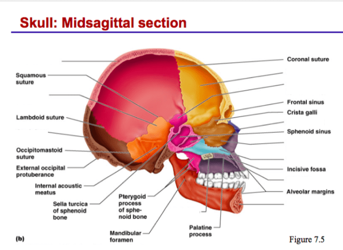
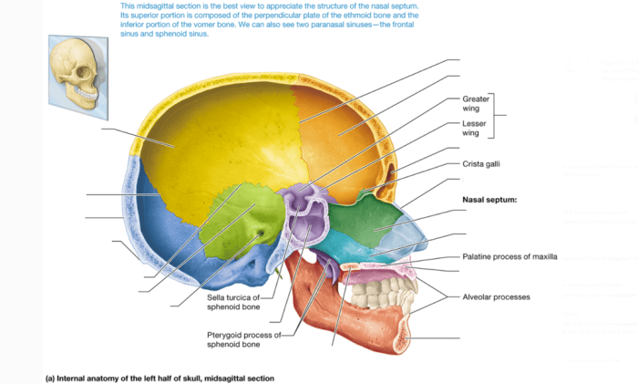
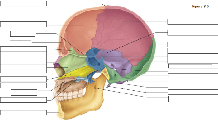
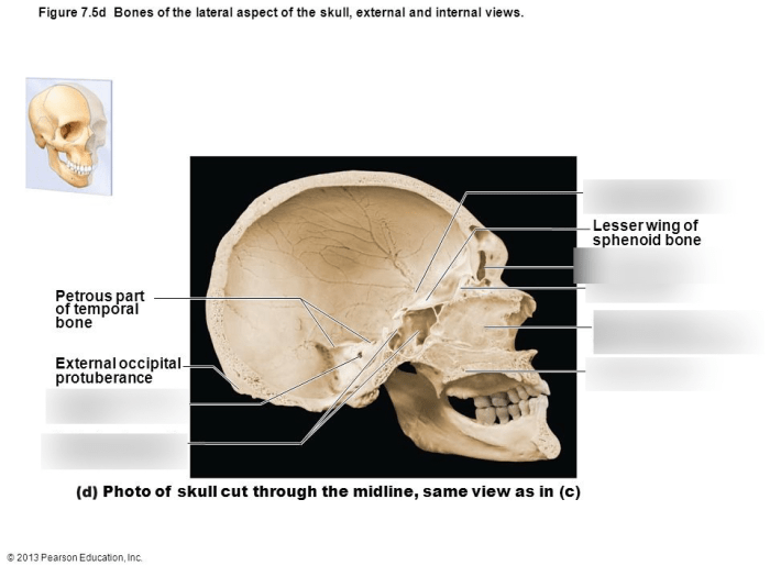
The internal midsagittal view of the skull offers a unique perspective on the intricate anatomy of the cranial cavity. This view, captured through advanced imaging techniques, provides invaluable insights for medical professionals, revealing the intricate relationships between various bony structures, midline structures, and potential skull pathologies.
This comprehensive guide delves into the anatomical landmarks, clinical significance, imaging techniques, anatomical variations, and surgical applications of the internal midsagittal view of the skull, providing a thorough understanding of its role in medical imaging and surgical interventions.
Internal Midsagittal View of the Skull

The internal midsagittal view of the skull provides a comprehensive view of the skull’s midline structures and their relationships. This view is essential for understanding the anatomy of the skull and diagnosing and assessing various skull pathologies.
Anatomical Landmarks
The internal midsagittal view of the skull shows the following bony structures:
- Frontal bone
- Parietal bones
- Occipital bone
- Ethmoid bone
- Sphenoid bone
- Maxillary bones
- Mandible
The midline structures visible in this view include:
- Sagittal suture
- Falx cerebri
- Tentorium cerebelli
- Pituitary gland
- Optic chiasm
- Basilar artery
Clinical Significance
The internal midsagittal view is a crucial view in medical imaging, particularly in:
- Neuroimaging: Assessing brain structures, detecting tumors, and diagnosing conditions like hydrocephalus.
- Dental imaging: Evaluating jaw structures, teeth, and dental implants.
- ENT imaging: Examining the nasal cavity, paranasal sinuses, and auditory structures.
Imaging Techniques, Internal midsagittal view of the skull
The internal midsagittal view can be obtained using various imaging modalities:
- Computed tomography (CT): Provides detailed cross-sectional images.
- Magnetic resonance imaging (MRI): Offers high-contrast images of soft tissues and bony structures.
- X-ray: Provides a basic overview of the skull’s bony structures.
Anatomical Variations
Normal anatomical variations in the internal midsagittal view include:
- Variations in the shape and size of the pituitary gland.
- Differences in the course of the basilar artery.
- Presence of accessory bones, such as the Inca bone.
Surgical Applications
The internal midsagittal view is used in neurosurgical procedures, including:
- Transcranial approaches to the brain.
- Endoscopic skull base surgery.
- Stereotactic radiosurgery.
Questions and Answers: Internal Midsagittal View Of The Skull
What is the internal midsagittal view of the skull?
The internal midsagittal view of the skull is an imaging plane that divides the skull into left and right halves and provides a detailed view of the midline structures of the cranial cavity.
What are the key anatomical landmarks visible in the internal midsagittal view?
The key anatomical landmarks visible in the internal midsagittal view include the frontal bone, ethmoid bone, sphenoid bone, sella turcica, pituitary gland, clivus, and occipital bone.
What is the clinical significance of the internal midsagittal view?
The internal midsagittal view is clinically significant in diagnosing and assessing various skull pathologies, including skull fractures, tumors, infections, and congenital anomalies.
What imaging techniques can be used to obtain an internal midsagittal view of the skull?
The internal midsagittal view of the skull can be obtained using various imaging techniques, including computed tomography (CT) and magnetic resonance imaging (MRI).
What are the surgical applications of the internal midsagittal view?
The internal midsagittal view is used in neurosurgical procedures to plan and guide surgical interventions, such as tumor resection, skull base surgery, and pituitary gland surgery.


